FYLA Supercontinuum Laser for Light Sheet Fluorescence Microscopy
2022-05-06
Advantages of elastic scattered light sheet fluorescence microscopy
Light Sheet Fluorescence Microscopy (LSFM) is a powerful imaging technique that enables fast and non-photo toxic 3D inspection of living specimens.
LSFM combines the speed of widefield imaging with moderate optical sectioning and low photobleaching. It is also referred to as SPIM, or simply “light sheet”. The defining feature of SPIM or LSFM is planar illumination of the focal plane from the side. Only a thin section of the sample is illuminated at any given time, minimizing photodamage and providing optical sectioning which improves SNR compared with widefield epifluorescence. Because the image is collected in a widefield (2D parallel) manner, light-sheet imaging is much faster than a point-scanned confocal microscope which detects only one pixel at a time.
It has become one of the most popular techniques for volumetric imaging because of three key features:
- Photodamage is minimized because excitation is confined near the focal plane, e.g. living things stay alive for much longer.
- Good optical sectioning is obtained, often approaching that of confocal microscopy.
- Acquisition rates are very fast, orders of magnitude faster than a traditional confocal microscope. The main disadvantage of SPIM is that extra optics are required to generate the light sheet.
Light Sheet microscopy is usually based on fluorescence techniques, and in general, the sample under study needs to be properly labeled to be imaged.
In this article, we will go deeper into this technique and study how elastically scattered light could be used to generate images of non-labeled samples.
The main obstacle is that these images are usually affected by speckle. To solve that inconvenience, they use an FYLA supercontinuum laser source of low temporal coherence FYLA supercontinuum source is presented as a candidate to reduce the speckle inherent in light-sheet microscopy images from scattered light.
Pablo Loza-Alvarez, Omar Alarte, David Merino of ICFO-Institut de Ciencies Fotoniques with Diego Battista and Giannis Zacharakis of Foundation for Research and Technology-Hellas use elastic scattered light from the sample to generate images, in order to avoid the need to label the sample.
In this work, they propose a novel light sheet based optical setup which implements three strategies for dealing with speckles of elastic scattering images:
- Polarization filtering
- Reducing the temporal coherence of the excitation laser light
- Reducing the spatial coherence of the light sheet.
These strategies enable pristine light-sheet elastic-scattering imaging of structural features in challenging biological samples avoiding the deleterious effects of speckle, and without relying on, but complementing fluorescent labeling.
Setup for polarization and coherence control in elastic scattering light sheet microscopy
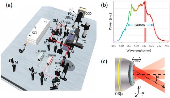
The main component of their elastic scattering light sheet microscope is the supercontinuum fiber laser from FYLA which emits a broadband spectrum of light from the visible to the infrared.
This source presents a very broad spectral bandwidth, at the same time, it presents very low temporal coherence. Both are desirable features in a light source when trying to reduce speckle effects in the images.
The FYLA white laser is used for Light sheet fluorescence microscopy selecting a band from 500 to 700 nm (140 nm FWHM), hence offering a lower temporal coherence for reduced speckle contrast.
Does the supercontinuum FYLA achieve label-free structural imaging in LSFM?
The results show that Iceblink FYLA supercontinuum source presents low speckle contribution on LSM images compared to other light sources with narrower spectrum bandwidth.
In conclusion, elastic scattering light sheet microscopy is a novel light sheet imaging modality suitable for label-free structural imaging.
In order to enhance the imaging quality in this configuration They have proposed to implement:
- Polarization control, which enables contrast selectivity and cancelling substrate background.
- Temporal and spatial coherence reduction, which enables extracting the endogenous intrinsic contrast from the speckle noise.
Implemented in this manner, elastic scattering light sheet imaging provides useful complementary structural information to standard LSFM experiments, as shown for MCTS samples. Also, it has the potential to provide relevant morphologic details of the samples, similar to histological sections, but in a non-destructive way.
Finally, elastic scattering light sheet microscopy is a promising technique which could enable new and interesting experiments, for example, as an alternative to LSFM in applications that are limited by low SNR, such as functional imaging or fast volumetric structural imaging.

Take a look of the entire paper: Enhanced Light Sheet Elastic Scattering Microscopy by Using a Supercontinuum Laser